Proceedings of the National Academy of Sciences Cover Image
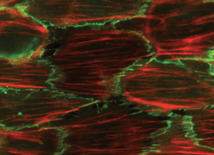
Actin stress fibers (red) orient perpendicular to the direction of cyclic stretch (horizontal) in arterial endothelial cells with normal Rho activity. Cell borders are indicated by B-catenin staining (green). The Rho pathway plays a major role in determining the direction and extent of stretch-induced stress fiber orientation.
Actin stress fibers in endothelial cells stretched at 1 Hz for 3 hours.
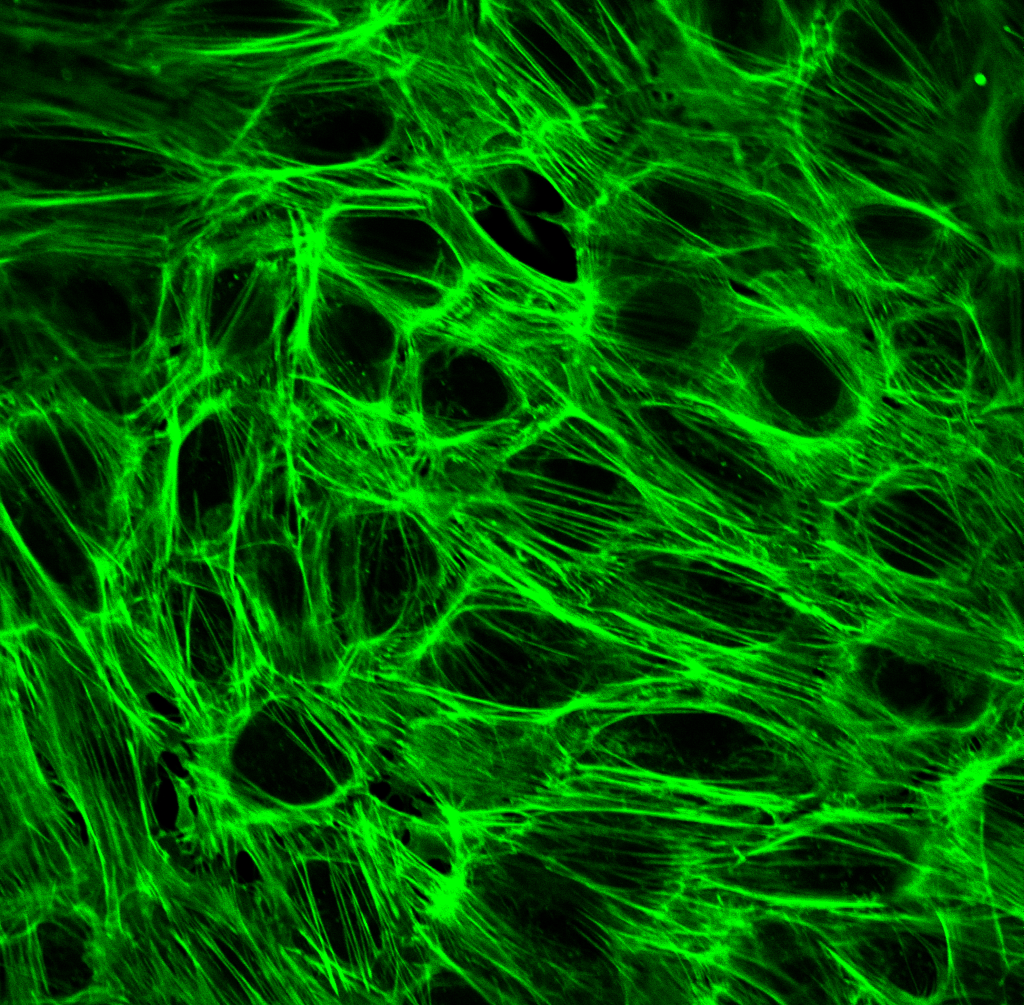
U2OS (osteosarcoma) cell expressing GFP-actin on a collagen hydrogel subjected to cyclic stretch.

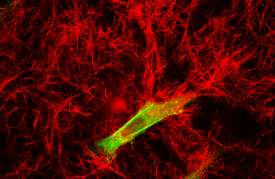
Cell-laden Collagen Microsphere Generation
Using microfluidics, droplets of collagen solution containing cells are polymerized to create monodisperse collagen microtissues. The micrograph shows a human mesenchymal stem cell and coolagen fibrils near the edge of a microsphere are shown.